Hip Fracture
Hip fractures can be very painful. For this reason, prompt surgical treatment is recommended. Treating the fracture and getting the patient out of bed as soon as possible will help prevent medical complications such as bed sores, blood clots, and pneumonia. In very old patients, prolonged bed rest can also lead to disorientation, which makes rehabilitation and recovery much more difficult.
Hip Fractures
Anatomy
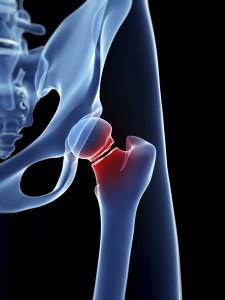
The hip is a ball-and-socket joint. The ball is the head of the femur, which is the upper part of the thighbone. The socket is called the acetabulum. The acetabulum is part of the pelvis bone. It has a rounded shape that fits around the femoral head.
Fractures of the acetabulum and pelvis are addressed in separate articles. Read more: Acetabular Fractures and Pelvic Fractures.
Description
A hip fracture can cause injury to one of four areas of the upper femur:
- Femoral neck. The area of the femur below the ball (femoral head).
- Intertrochanteric area. The area below the neck of the femur and above the long part or shaft of the femur. It is called intertrochanteric because it is marked by two bony landmarks: the greater trochanter and the lesser trochanter.
- Subtrochanteric area. The upper part of the shaft of the femur below the greater and lesser trochanters.
- Femoral head. The ball of the femur that sits in the socket.
Intertrochanteric and femoral neck fractures are the most common types of hip fracture. Femoral head fractures are extremely rare and are usually the result of a high-velocity event.
Cause
Most hip fractures result from low-energy falls in elderly patients who have weakened or osteoporotic bone. In these patients, even a simple twisting or tripping injury may lead to a fracture.
In some cases, the bone may be so weak that the fracture occurs spontaneously while someone is walking or standing. In this instance, it is often said that “the break occurs before the fall.” Spontaneous fractures usually occur in the femoral neck.
Stress fractures or fractures from repeated impact may also occur in the femoral neck. These fractures are often seen in long distance runners, particularly military recruits in basic training. When stress fractures occur in the subtrochanteric region of the hip, they are usually associated with prolonged use of certain osteoporosis medications.
Fractures of the femoral head are rare and are usually the result of a high-impact injury or are part of a fracture dislocation of the hip.
Symptoms
Typically, a hip fracture is acutely painful. The pain is usually localized to the groin and the upper part of the thigh. With most hip fractures, you will not be able to stand, bear weight, or move the upper part of your leg or knee. You will be able to move your ankle and toes, unless there is an injury to your lower leg in addition to your hip.
With some fractures, it may be possible to bear part of your weight on the leg—but it will be severely painful.
Doctor Examination
Physical Examination
Most of the time, a patient with a hip fracture will be taken by ambulance to a hospital emergency room.
At the hospital, you will be examined by either an emergency room physician or an orthopaedic surgeon. He or she will take a history of your injury and check to make sure that you do not have injuries to other parts of your body. The doctor will also check the sensation, movement, and circulation in your lower leg.
Often, the injured leg will appear shorter than the opposite leg and will be twisted or rotated, either internally or externally.
There may be a bruise on the outer part of the hip or thigh at the point of impact where you fell and all movement will be limited and painful.
A small number of hip fractures may not be as painful at first. Typically, these are nondisplaced fractures of the femoral neck. In this situation, you may choose to go to a doctor’s office rather than an emergency room. With this type of fracture, you may still be able to move your leg and bear weight even though it is painful.
Imaging Studies
Imaging studies will help confirm the diagnosis and provide more information about the fracture.
X-rays. X-rays provided images of dense structures, such as bone. Most hip fractures can be diagnosed with an x-ray.
Magnetic resonance imaging (MRI) scans. An MRI scan provides fine images of both soft tissue structures and bone. Because it is very sensitive, it can sometimes detect a small or incomplete fracture that cannot be seen on an x-ray.
Computerized tomography (CT) scans. A CT scan will provide a detailed cross-sectional image of your hip. Your doctor may order a CT scan to learn more about your fracture.
Treatment
This process of getting a patient medically ready for surgery is called optimization. Ideally, it should be done within 48 hours, but sometimes it may take longer. Even if it does takes longer, it is better to be optimized than to rush to surgery.
Treatment for a hip fracture depends upon the type and location of the fracture, as well as the age and condition of the patient.
Femoral Neck Fracture
This type of fracture is also sometimes called a subcapital or intracapsular fracture.
If a femoral neck fracture is not displaced, the most common treatment is in-situ pinning. In this procedure, surgical pins or screws are passed across the fracture site to hold the ball of the femur in place while the fracture heals. Pinning prevents the femoral head from dislodging or slipping off of the femoral neck, a situation that would require hip replacement.
A small number of femoral neck fractures may be treated without surgery. These are typically nondisplaced fractures in young healthy patients. Because there is a risk of displacement, they need to be monitored carefully. Nonoperative treatment consists of limited or protected weight bearing with crutches for several weeks.
Patients who did not walk before surgery or have severe medical problems may also be considered for nonsurgical treatment.
Displaced fractures of the femoral neck present a more difficult problem than nondisplaced fractures. The blood supply to the head of the femur comes through a structure called the posterior capsule. The posterior capsule is often injured with a displaced femoral neck fracture, so the fracture has less chance of healing.
Even if the fracture does heal, a condition called avascular necrosis may develop in the head of the femur. This causes damage to the bone cells, collapse of part of the femoral head, and subsequent arthritis.
For this reason, displaced fractures of the femoral neck are often treated with hip replacement. For elderly patients, a hemi-arthroplasty, or partial hip replacement, is the treatment of choice. In younger, more active patients, total hip replacement may be considered.
In some younger patients, it is desirable to preserve the natural femoral head rather than do a replacement. In these patients an open reduction is performed. This involves making an incision over the hip joint, putting the bone back into place, and then holding the bone with pins, screws, or another type of metal fixation. While this procedure does carry the risks of avascular necrosis and failure to heal, it is felt best to try to preserve a younger person’s normal hip.
Your orthopedic surgeon will review the treatment options with you and your family.
Intertrochanteric Fracture
Intertrochanteric fractures occur below the femoral neck in a broader region between the greater and lesser trochanters. The greater trochanter is the bump you feel on the side of your hip. There may be two, three, or even multiple fracture fragments.
Intertrochanteric fractures are treated surgically with either a sliding compression hip screw and side plate or an intramedullary nail.
The compression hip screw is fixed to the outer side of the bone with bone screws. A large secondary screw (lag screw) is placed through the plate into the femoral head and neck. This design allows for impaction and compression at the fracture site. This will increase stability and promote healing.
The intramedullary nail is placed directly into the marrow canal of the bone through an opening made at the top of the greater trochanter. One or multiple screws are then placed through the nail and into the femoral head.
Fracture of the Greater Trochanter
Isolated fractures of the greater trochanter usually come from a low-energy household fall. While they are often painful, they usually heal without surgery. These fractures are stable and can be treated with protected weight bearing with either crutches or a walker.
If an x-ray shows an isolated fracture of the greater trochanter, it is often helpful to obtain an MRI to make sure that the fracture does not extend to the intertrochanteric area.
Subtrochanteric Fracture
Subtrochanteric fractures involve the upper part of the shaft of the femur, just below the hip joint.
They are treated surgically with an intramedullary nail into the shaft of the femur and a screw placed through the nail into the femoral head.
To keep the bones from rotating around the nail or from shortening (telescoping) on the nail, additional screws may be placed at the lower end of the nail near the knee. These are called interlocking screws.
In some cases, your surgeon may choose to use a compression screw with a long side plate instead of a nail.
Femoral Head Fracture
Femoral head fractures are rare; they account for less than 1 percent of all hip fractures. They usually result from a high-velocity event. Sometimes there may be an associated fracture of the hip joint socket.
If the fracture is not displaced, it may be treated nonsurgically with limited weight bearing. If there is a small displaced fragment that does not involve a large part of the joint surface, then the fragment may be simply removed.
If there is a large fragment in a young active person, open reduction and fixation with screws is often done. In an older person, hip replacement—either partial or total—to replace the damaged femoral head is the treatment of choice.
Recovery
Most patients are able to get of bed and start physical therapy the day after surgery. It is important to begin moving as soon as possible. This helps prevents medical complications, such as blood clots, pneumonia, and bed sores. For older patients, it also helps prevent disorientation and deconditioning.
Hip fractures in the elderly may cause disability and lack of independence. Early movement and rehabilitation have been shown to improve long-term results.
During recovery, you will work with physical and occupational therapists. They will give you exercises and tell you how much weight you can put on your leg. They will also show you how to manage the activities of daily living, such as bathing and dressing.
Medical Care
If you take medications for a problem such as heart disease, an internist or hospitalist will most likely assist in your care. He or she will address any medical problems and prescribe the appropriate treatment.
You will be given antibiotics to prevent infection for 24 hours after surgery.
You will also be placed on a blood thinner to help prevent blood clots in your legs. This may be given as either pills or injections. Your doctor will determine the length of time you need to be on blood thinners. Compression boots may be placed on your legs while you are in bed.
Pain Management
Your doctor and nurses will work with you to help reduce your pain. Typically, you will be given medicine through an IV in the first few hours after surgery. Then you will be switched to oral pain medicine. This may take the form of opioids, nonsteroidal anti-inflammatories, or over-the-counter pain medication such as Tylenol. Your doctor may also use a combination of these medications to manage pain.
Be aware that, while opioids can relieve pain after surgery, they are a narcotic and can be addictive. You should stop taking opioids as soon as your pain begins to improve. Your surgeon will help you with pain management during your recovery.
Rehabilitation
Many patients go home after hip fracture surgery, but some will need short-term care in a rehabilitation facility. Usually, these patients are elderly or have no caregivers at home.
If you go to a rehabilitation facility, you will need to stay there until you can walk independently and manage your daily activities.
If you go directly home, you will have in-home physical therapy until you are strong enough to go to therapy at an outside facility.
Your doctor will manage your care in the postoperative period. He or she will check the wound, prescribe physical therapy, and take x-rays to monitor the healing.
If you have had a procedure that includes internal fixation, it may be several weeks until you can bear full weight. If you have had a hip replacement, you can most likely be fully weight bearing right away.


There are no reviews yet.